Belying their placid illustrations in textbooks, living biological cells are bustling hubs of activity where substances constantly traffic in and out. Passage through a cell’s encompassing membrane, however, is no simple feat; the process typically involves dozens of proteins precisely interacting, all in the right places and at the right times.
Unraveling this process and other subcellular marvels is a chief research aim of Margaret Johnson’s lab. She and her colleagues broadly study dynamical systems in biology, seeking to mathematically quantify how large molecules—so-called macromolecules—naturally organize into the complex rudiments for life.
In this way, the research is a perfect amalgam of Johnson’s academic interests. “Math was always my favorite subject, but I also loved animals. Biology and the study of living systems was the most fascinating part of science to me,” says Johnson, who joined Johns Hopkins in 2013 and is now an associate professor in the Thomas C. Jenkins Department of Biophysics. “In my lab, we use mathematical modeling and computer simulations to answer unresolved questions about how, why, and how well macromolecular self-assembly is controlled in living systems.”
courtesy of the Johnson Lab
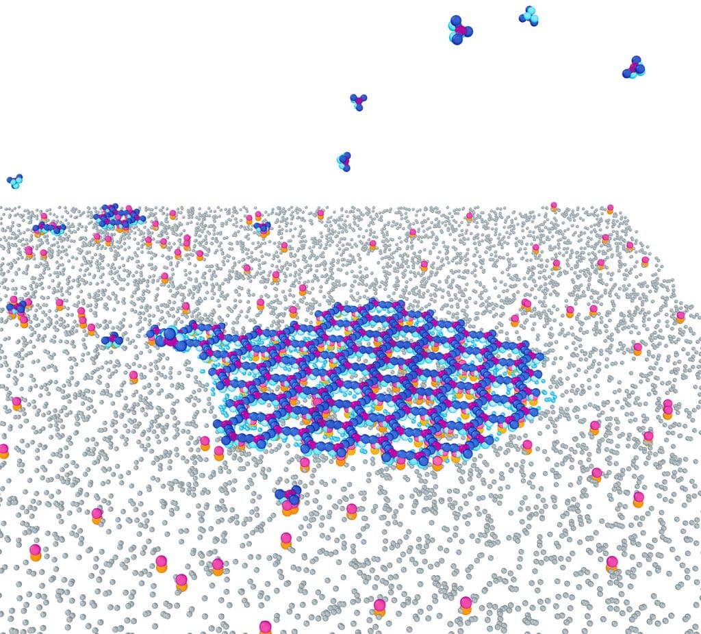
Above, clathrin recruitment and assembly on membranes through physics-based modeling.
How materials get into cells
One specialty area for Johnson’s group is endocytosis, the process whereby materials are transported into cells. The researchers are probing the mechanisms of clathrin-mediated endocytosis, a major subtype involving a protein called clathrin that helps form cage-like spheres around the cell membrane to bring cargo-bound receptors into the cell.
Gaining a better understanding of how the networks of proteins involved in this transport mode come together could lead to new insights into a vast range of conditions and diseases. To rattle off just a few of its roles, clathrin-mediated endocytosis ferries essential nutrients into cells, reloads nerves for continuous signal-sending, and (unwittingly) enables invading viruses to gain cellular entry.
In my lab, we use mathematical modeling and computer simulations to answer unresolved questions about how, why, and how well macromolecular self-assembly is controlled in living systems.”
—Margaret Johnson
Getting at the nitty-gritty of this kind of endocytosis has required Johnson and colleagues to create new computer models that simulate the physics and chemistry at play. “Existing models are not capable of addressing many of the questions about the spatial and temporal dynamics that control self-assembly at the cell scale,” says Johnson.
Building on the results
Using these techniques, the research group recently revealed why clathrin structures often form but then spontaneously disassemble before transporting anything into a cell. Other kinds of proteins that link clathrin to the cell membrane and to cargo-bound receptors must be present in sufficient quantities, the study predicts, thereby linking the assembly success to the membrane composition and offering guidance for future experiments.
The team is building off those results, not only in clathrin-mediated endocytosis, but other cellular processes involving self-assembly. One example is viral budding, where viral proteins produced by our own cell’s machinery will assemble within infected cells to exit through the cell membrane and go on to newly infect additional cells. Johnson and colleagues are studying how the viral components have evolved to ensure productive assembly, budding, packaging of the viral genetic information, and maturation (for HIV) into fully infectious virions.
“The tools we make to learn about endocytosis are also applicable to diverse pathways in cellular biology,” says Johnson. “Predicting how these essential processes are controlled in complex environments is critical to understanding their function in healthy, stressed, or diseased cells.”